Pharmacological models to assess the role of tryptophan-metabolizing enzymes in cancer

Cell-based and co-culture assays, tumor mouse models, and screening in primary patient material

  • L-tryptophan is a key regulator of the immune modulatory activity of T cells and natural killer cells.
  • A comprehensive set of pharmacological models has been applied to study the role and inhibition of the L-tryptophan-catabolizing enzyme IDO1, an important target for cancer immunotherapy.

  • Decreased IDO1 activity was observed after incubation of the enzyme or IDO1-expressing cells with the inhibitors epacadostat and NTRC 3883-0, using the NFK GreenScreen™ assay technology.
  • Co-culture of IDO1-overexpressing cells with immune cells showed restoration of cytotoxic T cell proliferation by epacadostat and NTRC 3883-0.

  • Two syngeneic mouse models were applied for the independent assessment of the efficacy of epacadostat: the colon carcinoma CT26 model and a melanoma model that uses IDO1-overexpressing B16F10 cells.

  • The IDO1-overexpressing cell line was generated at NTRC. The in vivo experiments were carried out at CROs. Compound and metabolite levels in plasma, tumors and normal tissues were analyzed by LC-MS/MS.

  • For the first time, the use of primary cell cultures isolated from ascites of ovarian cancer patients was demonstrated for the determination of tumor IDO1 expression and activity.

  • The different in vitro and in vivo models provide an extensive pharmacological characterization of inhibitors for cancer immunotherapy and support further development of inhibitors with differentiating properties.

Paper Frontiers In Immunology Co Cultures
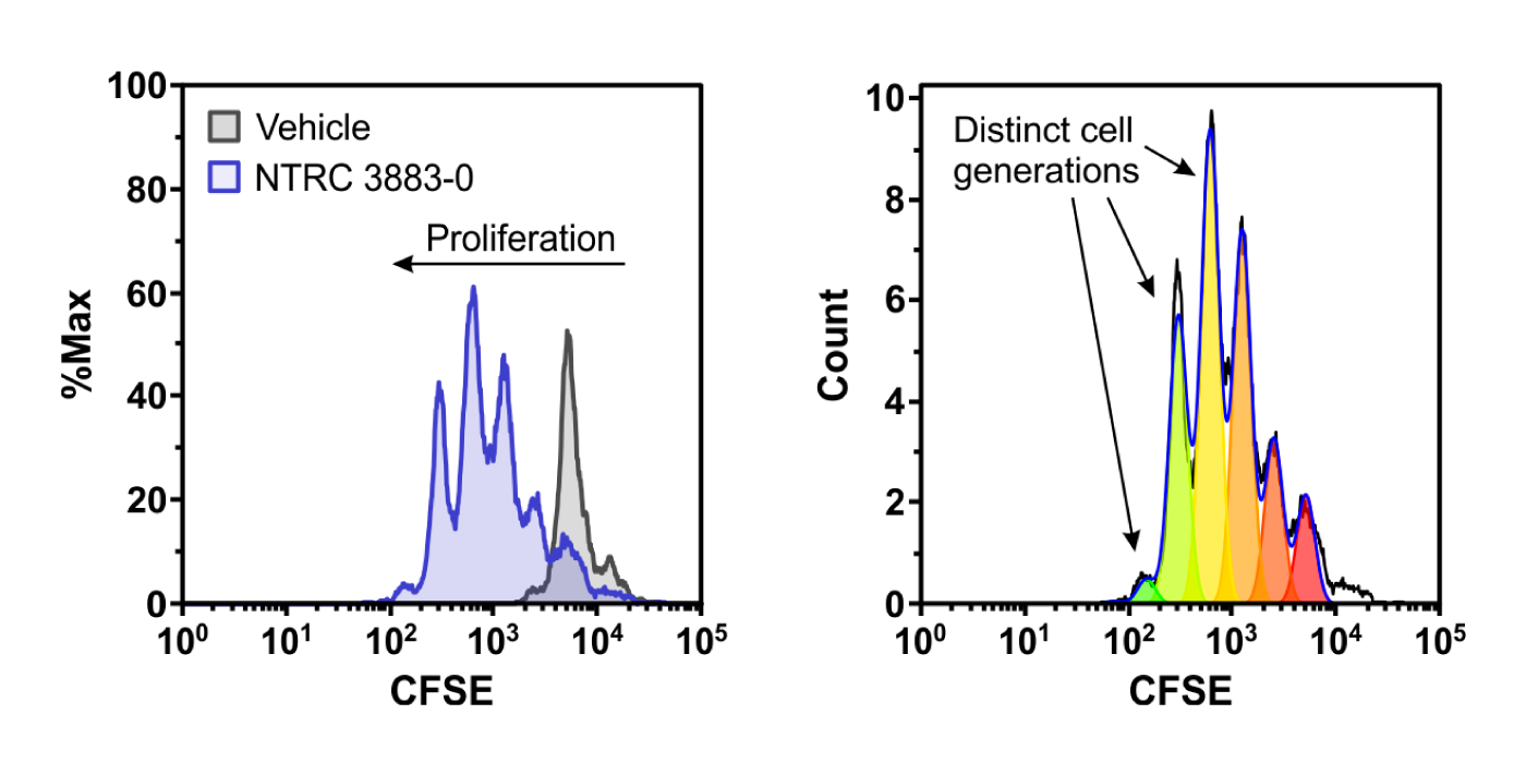
Co-culture assay of IDO1-overexpressing HEK-293 cells with lymphocytes from a healthy donor. IDO1 inhibitor NTRC 3883-0 restores proliferation of the fluorescently labeled CD8-positive T cells (left), as indicated by the multiple cell generations present in the T cell population (right).
Paper Frontiers In Immunology Fig 5 IDOTDO
Illustration: Expression and modulation of IDO1 and TDO in cell cultures isolated from ovarian cancer ascites. The left chart shows a tukey boxplot of basal IDO1 and TDO2 gene expression as determined by qPCR in early passage cell cultures. The right chart shows inhibition of the Trp-catabolizing activity in IFNγ-stimulated samples by NTRC 3883-0 (closed symbols) and epacadostat (open symbols).